Distinct Biological Activity of Lipopolysaccharides with Different Lipid A Acylation Status from Mutant Strains of Yersinia pestis and Some Members of Genus Psychrobacter
K. V. Korneev1,2,3, A. N. Kondakova4, N. P. Arbatsky4, K. A. Novototskaya-Vlasova5, E. M. Rivkina5, A. P. Anisimov6, A. A. Kruglov2,3, D. V. Kuprash1,2,3, S. A. Nedospasov1,2,3, Yu. A. Knirel4*#, and M. S. Drutskaya1*#
1Engelhardt Institute of Molecular Biology, Russian Academy of Sciences, ul. Vavilova 32, 119991 Moscow, Russia; fax: (499) 135-1405; E-mail: marinadru@gmail.com2Department of Immunology, Biological Faculty, Lomonosov Moscow State University, 119991 Moscow, Russia
3Belozersky Institute of Physico-Chemical Biology, Lomonosov Moscow State University, 119991 Moscow, Russia
4Zelinsky Institute of Organic Chemistry, Russian Academy of Sciences, Leninsky pr. 47, 119991 Moscow, Russia; fax: (499) 135-5328; E-mail: yknirel@gmail.com
5Institute for Physicochemical and Biological Problems in Soil Science, Russian Academy of Sciences, 142290 Pushchino, Moscow Region, Russia; fax: (4967) 33-0532; E-mail: soil@issp.serpukhov.su
6State Research Center for Applied Microbiology and Biotechnology, 142279 Obolensk, Moscow Region, Russia; fax: (4967) 36-0010; E-mail: info@obolensk.org
* To whom correspondence should be addressed.
# These authors contributed equally to this work.
Received June 30, 2014
Correlation between the chemical structure of lipid A from various Gram-negative bacteria and biological activity of their lipopolysaccharide (LPS) as an agonist of the innate immune receptor Toll-like receptor 4 was investigated. Purified LPS species were quantitatively evaluated by their ability to activate the production of tumor necrosis factor (TNF) by murine bone marrow-derived macrophages in vitro. Wild-type LPS from plague-causing bacteria Yersinia pestis was compared to LPS from mutant strains with defects in acyltransferase genes (lpxM, lpxP) responsible for the attachment of secondary fatty acid residues (12:0 and 16:1) to lipid A. Lipid A of Y. pestis double ΔlpxM/ΔlpxP mutant was found to have the chemical structure that was predicted based on the known functions of the respective acyltransferases. The structures of lipid A from two members of the ancient psychrotrophic bacteria of the genus Psychrobacter were established for the first time, and biological activity of LPS from these bacteria containing lipid A fatty acids with shorter acyl chains (C10-C12) than those in lipid A from LPS of Y. pestis or E. coli (C12-C16) was determined. The data revealed a correlation between the ability of LPS to activate TNF production by bone marrow-derived macrophages with the number and the length of acyl chains within lipid A.
KEY WORDS: lipopolysaccharide, lipid A, Toll-like receptor 4, tumor necrosis factor, Yersinia pestis, Psychrobacter spp.DOI: 10.1134/S0006297914120062
Lipopolysaccharide (LPS) of most Gram-negative bacteria induces intracellular signaling through the innate immune receptor, Toll-like receptor 4 (TLR4) [1]. Although the three-dimensional structure of the LPS–MD-2–TLR4 complex was established by X-ray analysis [2], some aspects of structure–function relationships for various LPS are not fully understood. Currently, the best-studied TLR4 agonists are a highly active hexaacyl lipid A from Escherichia coli and its tetraacyl biosynthetic precursor, lipid IVa, which is a weak activator [3, 4]. However, the natural diversity of ligands with potentially different lipid A structures is certainly not limited to the studied structures. Therefore, investigation of “non-canonical” forms of LPS, primarily those that differ in the structure of the lipid A moiety, which is largely responsible for innate immune recognition, is of interest.
The “canonical” hexaacyl lipid A consists of two phosphorylated glucosamine residues with four primary hydroxylated acyl groups attached (R2, R3, R2′, R3′), two of which carry secondary non-hydroxylated acyl residues (R2′′, R3′′) [5]. It should be noted that five acyl groups of lipid A (R3, R2′, R2′′, R3′, R3′′) are docked in a hydrophobic pocket of the adapter protein MD-2 formed by two β-sheets, rather than interacting with TLR4 directly, whereas the glucosamine residues remain on the surface [6]. The R2 acyl group is bound by hydrophobic interactions to the neighboring TLR4 monomer rather than to MD-2. The binding causes conformational changes in TLR4, activation of its intracellular domain, and subsequent triggering of intracellular signaling pathways [7, 8]. The resulting activation of the transcription factor NF-κB induces transcription of several proinflammatory cytokine genes including tnf encoding for TNF (tumor necrosis factor), the major factor in septic shock [9, 10].
In the present study, we examined correlation between the chemical structure of LPS from Gram-negative bacteria, namely the length and the number of the acyl chains in lipid A, and the biological activity of LPS as a TLR4 agonist.
MATERIALS AND METHODS
Bacterial cultures. The following strains of Gram-negative bacteria were used: Escherichia coli O130, Francisella tularensis 15, Yersinia pestis KM260(11) and its mutants KM260(11)ΔlpxM, KM260(11)ΔlpxP, KM260(11)ΔlpxM/ΔlpxP [11, 12], Psychrobacter arcticus DSM 17307T, and Psychrobacter cryohalolentis DSM 17306T [13]. Bacteria of the genus Psychrobacter were obtained from the All-Russian Collection of Microorganisms.
Strains of P. cryohalolentis and P. arcticus were grown at 24°C, Y. pestis at 25°C, and E. coli and F. tularensis at 37°C in a 10-liter fermenter (New Brunswick Scientific Fermentors, USA) in a liquid aerated medium until the initial stage of the stationary phase.
Strains of Y. pestis, E. coli, and F. tularensis were grown at pH 6.9-7.1 in a medium that contained (per liter of distilled water) 20-30 g pancreatic fish-flour hydrolysate, 10 g yeast autolysate, 3.9 g glucose, 6 g K2HPO4, 3 g KH2PO4, and 0.5 g MgSO4. After 48-h incubation, bacterial biomass was centrifuged, frozen at –70°C, and lyophilized. A solid agar medium was prepared by adding 2% agar to the liquid medium.
Strains of P. cryohalolentis and P. arcticus were grown at pH 7.6 in a medium that contained (per liter of distilled water) 4 g yeast extract, 1.12 g Na2HPO4, 0.4 g KH2PO4, 19.5 g NaCl, 2 g NH4Cl, 4 g MgSO4, 0.1 ml CaCl2 (10%), 0.05 ml FeSO4 (1%), and 10 ml microelement solution SL-10 (990 ml dH2O, 10 ml HCl (25% 7.7 mM), 1.5 g FeCl2·4H2O, 70 mg ZnCl2, 100 mg MgCl2·4H2O, 6 mg H3BO3, 190 mg CoCl2·6H2O, 24 mg NiCl2·6H2O, 36 mg NaMoO4·2H2O). The biomass was dried with acetone according to the standard protocol [14].
Isolation of LPS. LPS was isolated from dried bacterial cells of each strain by extraction with a mixture of phenol, chloroform, and petroleum ether at 25°C and purified by ultracentrifugation at 105,000g (twice) followed by treatment with DNase, RNase, and proteinase K to remove nucleic acids and proteins. Highly purified LPS preparations were obtained by hydrophobic gel chromatography in sodium acetate buffer in the presence of EDTA and sodium deoxycholate controlled by UV monitoring and polyacrylamide gel electrophoresis. Two approaches were used: 1) an LPS sample was fractionated by gel chromatography on Sephadex G-150, the LPS-containing fraction was lyophilized, an excess of salts was removed by extraction with ethanol, the precipitate was dissolved in water, and LPS was precipitated with ethanol and lyophilized; 2) an LPS sample was fractionated on AcA 44 Ultragel, the LPS-containing fractions were pooled and dialyzed first against 0.2% NaHCO3, then against distilled water and lyophilized. The purified LPS preparations obtained by both methods were free from proteins and nucleic acids. Lipid A was cleaved by mild acid hydrolysis of LPS, the lipid precipitate was suspended in water, lyophilized, and the dried preparation was extracted with chloroform–methanol (1 : 1 v/v) to remove phospholipid contaminations.
Mass spectrometry of LPS. LPS was subjected to mild acid hydrolysis with 2% AcOH at 100°C until precipitation of lipid A (2-4 h). The resulting precipitate was separated by centrifugation and lyophilized. Mass spectra were recorded on a Bruker ApexII spectrometer (Bruker Daltonics, USA) with electrospray ionization and ion detection using Fourier transform ion-cyclotron resonance [15]. Negative ion spectra were recorded and processed using standard experimental sequences as provided by the manufacturer. Mass accuracy was checked through external calibration. Samples (10 ng/ml) were dissolved in a mixture of isopropanol, water, and triethylamine (50 : 50 : 0.001 v/v) and sprayed at a flow rate of 2 µl/min. The capillary output voltage was set to 3.8 kV, and the drying gas temperature was 150°C.
Laboratory animals. C57Bl/6 mice and tlr4-deficient mice (A. A. Kruglov et al., manuscript in preparation) 8-10-week-old (weight ~20 g) were used. The mice were housed in the Pushchino Animal Breeding Facility of the Pushchino branch of Shemyakin and Ovchinnikov Institute of Bioorganic Chemistry, Russian Academy of Sciences, under specific pathogen free conditions.
Bone marrow-derived macrophage cultures and activation. Primary bone marrow-derived macrophage (BMDM) cultures were obtained by in vitro differentiation of bone marrow cells from C57Bl/6 mice or TLR4-deficient mice. The cells were maintained for 10 days in a culture medium according to the standard protocol [16]. The medium for BMDM cultures contained DMEM (Gibco, USA) with 2 mM L-glutamine (HyClone, USA) supplemented with 20% horse serum (HyClone) and 30% conditioned medium from L929 cells (source of M-CSF). For measuring TNF concentration in the supernatants, primary bone marrow-derived macrophages were plated on 96-well plates (5·104 cells per well). DMEM medium without serum that contained 1 or 10 ng/ml LPS was added, and after 4-h incubation in a CO2-incubator at 37°C, the supernatant was transferred to a 96-well plate and stored at –80°C for further analysis.
Analysis of TNF production. The amount of TNF produced in the cell supernatant was determined by ELISA using a Mouse TNFalpha ELISA Ready-SET-Go kit (eBioscience, USA) according the manufacturer’s protocol.
Statistical analysis. The GraphPad Prism 6 program (GraphPad Software) was used for statistical data analysis. The Mann–Whitney U-test was used for nonparametric comparison of two independent data samples and determination of the degree of reliability. The data were obtained in three independent experiments and presented as the mean ± SD for TNF-α concentration in the supernatant. Differences between the groups were considered significant at P < 0.05.
RESULTS AND DISCUSSION
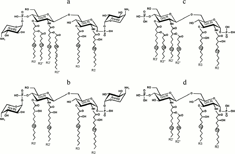
Structures of lipid A in wild-type LPS of Y. pestis KM260(11) [15] and its mutants in genes lpxM [11, 12] and lpxP [12] encoding lauroyl (12:0) transferase and palmitoleoyl (16:1) transferase, respectively, were established earlier. The wild-type lipid A synthesized at 25°C has six acyl groups, from which four 3-hydroxymyristoyl groups (3HO14:0) are primary (i.e. attached directly to the glucosamine residues) and two other groups (12:0 and 16:1) are secondary (Fig. 1a). Lipid A structures of KM260(11)ΔlpxM and KM260(11)ΔlpxP mutants lack the secondary lauroyl 12:0 group (R3′′) or palmitoleoyl 16:1 group (R2′′), respectively, and thus both represent a pentaacyl lipid A. Structure of lipid A of the double KM260(11)ΔlpxM/ΔlpxP mutant was elucidated by mass spectrometry in this study. As predicted from the genetic data, this mutant lacks both secondary acyl groups, 12:0 and 16:1, and is thus represented by the tetraacyl form (Fig. 1b).
Fig. 1. Structures of hexaacyl and tetraacyl bisphosphoryl lipids A in LPS of wild-type Y. pestis and its ΔlpxM/ΔlpxP mutant grown at 25°C (a, b) and Psychrobacter spp. (c, d). GlcN, glucosamine; Ara4N, 4-amino-4-deoxyarabinose; R2, R3, R2′, R3′, primary acyl groups; R2′′ and R3′′, secondary acyl groups. Numbers indicate the number of carbon atoms in the acyl chain; in case of Psychrobacter spp., the numbers refer to the predominant form of lipid A.
In addition, lipid A of the two representative psychrotrophic bacteria of the genus Psychrobacter was characterized chemically for the first time. As judged by mass spectrometry data, it includes fatty acids with relatively short acyl chains (Fig. 1c). The predominant hexaacyl form has four primary 3HO12:0 groups and two secondary 10:0 groups attached to the primary fatty acids. There is also tetraacyl lipid A that lacks one residue each of 3HO12:0 and 10:0 (Fig. 1d), and a small amount of pentaacyl lipid A. The main sources of structural heterogeneity are: a) the presence of monophosphoryl lipid A in addition to the major bisphosphoryl lipid A, and b) a different length of acyl chains resulting in the presence of minor species in which the primary 3HO12:0 group is replaced with a 3HO10:0 group and one or both secondary 10:0 groups are replaced with 12:0 or 11:0. The relative percentage of various forms of lipid A in LPS of Psychrobacter spp. is summarized in the table.
Total relative percentage of various forms of lipid A in LPS of
Psychrobacter spp. (based on mass spectral data of isolated
lipids A) and calculated (Mcalc) and experimental
(Mexp) molecular masses of the predominant forms of lipid A
(Da)

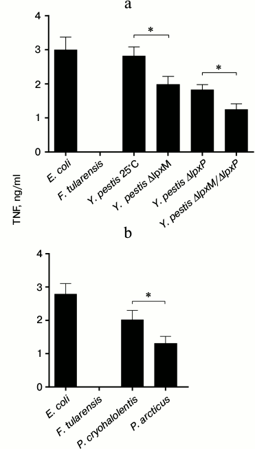
LPS of E. coli with highly active hexaacyl lipid A was used as positive control for assessing TNF production in activated macrophages [5]. LPS of F. tularensis with tetraacyl monophosphoryl lipid A, which does not induce TLR4 signaling [17, 18], was used as a negative control.
To evaluate the specificity of TLR4 signaling, the LPS preparations were also tested in bone marrow macrophage cultures deficient in TLR4. TNF production by TLR4-deficient macrophages in response to LPS did not exceed the detection limit of the method (data not shown). This observation confirmed the specificity of our LPS preparations and, therefore, TNF production by BMDM is triggered by the interaction of LPS with TLR4.
BMDM from wild-type C57Bl/6 mice were activated by LPS samples from Y. pestis and Psychrobacter spp. at various concentrations (0.1, 1, 10, and 100 ng/ml) to determine the optimal working concentration of LPS (data not shown). For LPS from Y. pestis mutants, the most marked differences in TNF induction were observed at 1 ng/ml, and for LPS from Psychrobacter spp. at 10 ng/ml. The higher concentration in the latter case can be accounted for by the presence of a long-chain polysaccharide in the S-form LPS of Psychrobacter spp. [19], which decreased the content of lipid A in LPS, whereas there is no such component in the R-form LPS of Y. pestis mutants [15].
Figure 2a shows that wild-type LPS of Y. pestis grown at 25°C activated TNF production approximately as efficiently as LPS of E. coli, which is consistent with the presence of active hexaacyl lipid A in both strains. In Y. pestis mutants, the absence of at least one acyl group was crucial for binding of LPS to TLR4. LPS from ΔlpxM and ΔlpxP mutants induced TNF production at approximately the same level, the former being a slightly stronger activator than the latter. Although this difference did not reach the level of statistical significance, it indicated that the absence of the 16:1 acyl group (R2′′) might be more important for manifestation of the biological activity of lipid A than the absence of the 12:0 group (R3′′). This effect may be due to a better anchoring of the LPS molecule into the hydrophobic pocket of the MD-2 adapter protein provided by the longer-chain 16:1 group with a double carbon–carbon bond than by the shorter-chain saturated 12:0 group. As expected, LPS of double ΔlpxM/ΔlpxP mutant induced the weakest TNF production by macrophages. Therefore, the biological activity of Y. pestis LPS depends on the number of acyl groups in the lipid A moiety.
Fig. 2. TNF production in bone marrow-derived macrophage cultures in response to LPS from various strains of Y. pestis at 1 ng/ml (a) and Psychrobacter spp. at 10 ng/ml (b). * P < 0.05. The values for LPS of F. tularensis were below the detection limit.
A comparison of the biological activity of LPS from strains of Psychrobacter spp. revealed the following (Fig. 2b): LPS of P. cryohalolentis with hexaacyl lipid A as the predominant form induced the strongest TNF production; LPS P. arcticus was a weaker activator that correlated with a lower content of hexaacyl lipid A and, accordingly, a higher content of pentaacyl and tetraacyl forms (table).
Therefore, this study demonstrated the functional importance of the number of acyl groups in lipid A, which correlated with the observed ability of LPS to activate TNF production in primary bone marrow-derived macrophages. A lower level of activation of TNF production by macrophages in response to LPS from strains of Psychrobacter spp. in general can be accounted for by shorter primary and secondary acyl groups in their lipids A (C10-C12) compared with Y. pestis and E. coli lipids A (C12-C16), which presumably do not provide an equally efficient LPS binding to the MD-2–TLR4 complex. It can be concluded that the total contribution of the acyl groups in lipid A impacts directly the biological activity of LPS: the activity increases with an increase in the number and the chain length of the acyl groups.
Authors thank S. V. Dentovskaya, S. A. Ivanov, and R. Z. Shaikhutdinova for providing LPS of Yersinia pestis mutants, B. Lindner (Research Center Borstel, Borstel, Germany) for providing access to a Bruker ApexII mass spectrometer, Head of the Laboratory of Anaerobic Microorganisms of IBPM RAS V. A. Shcherbakova for providing strains of Psychrobacter spp., R. V. Zvartsev for mouse genotyping, A. A. Chashchina and E. N. Sviryaeva for help with performing some experiments, and Head of the Pushchino Animal Breeding Facility of the Pushchino branch of the Institute of Bioorganic Chemistry G. B. Telegin for breeding and providing mice for the experiments.
This work was supported by the Russian Foundation for Basic Research (grants 13-04-40266 for D. B. Kuprash, 13-04-40269 for Yu. A. Knirel, 13-04-40268 for A. A. Kruglov, and 10-04-01470 for M. S. Drutskaya).
REFERENCES
1.Poltorak, A., He, X., Smirnova, I., Liu, M. Y., Van
Huffel, C., Du, X., Birdwell, D., Alejos, E., Silva, M., Galanos, C.,
Freudenberg, M., Ricciardi-Castagnoli, P., Layton, B., and Beutler, B.
(1998) Defective LPS signaling in C3H/HeJ and C57BL/10ScCr mice:
mutations in Tlr4 gene, Science, 282,
2085-2088.
2.Park, B. S., Song, D. H., Kim, H. M., Choi, B. S.,
Lee, H., and Lee, J. O. (2009) The structural basis of
lipopolysaccharide recognition by the TLR4-MD-2 complex, Nature,
458, 1191-1195.
3.Kovach, N. L., Yee, E., Munford, R. S., Raetz, C.
R., and Harlan, J. M. (1990) Lipid IVA inhibits synthesis and release
of tumor necrosis factor induced by lipopolysaccharide in human whole
blood ex vivo, J. Exp. Med., 172, 77-84.
4.Golenbock, D. T., Hampton, R. Y., Qureshi, N.,
Takayama, K., and Raetz, C. R. (1991) Lipid A-like molecules that
antagonize the effects of endotoxins on human monocytes, J. Biol.
Chem., 266, 19490-19498.
5.Qureshi, N., Takayama, K., Mascagni, P., Honovich,
J., Wong, R., and Cotter, R. J. (1988) Complete structural
determination of lipopolysaccharide obtained from deep rough mutant of
Escherichia coli. Purification by high performance liquid
chromatography and direct analysis by plasma desorption mass
spectrometry, J. Biol. Chem., 263, 11971-11976.
6.Lu, Y. C., Yeh, W. C., and Ohashi, P. S. (2008)
LPS/TLR4 signal transduction pathway, Cytokine, 42,
145-151.
7.Kawai, T., and Akira, S. (2010) The role of
pattern-recognition receptors in innate immunity: update on Toll-like
receptors, Nat. Immunol., 11, 373-384.
8.Maeshima, N., and Fernandez, R. C. (2012)
Recognition of lipid A variants by the TLR4–MD-2 receptor
complex, Front. Cell. Infect. Microbiol., 3, 3.
9.Glauser, M. P., Zanetti, G., Baumgartner, J. D.,
and Cohen, J. (1991) Septic shock: pathogenesis, Lancet,
338, 732-736.
10.Chow, J. C., Young, D. W., Golenbock, D. T.,
Christ, W. J., and Gusovsky, F. (1999) Toll-like receptor-4 mediates
lipopolysaccharide-induced signal transduction, J. Biol. Chem.,
274, 10689-10692.
11.Anisimov, A. P., Shaikhutdinova, R. Z.,
Pan’kina, L. N., Feodorova, V. A., Savostina, E. P., Bystrova, O.
V., Lindner, B., Mokrievich, A. N., Bakhteeva, I. V., Titareva, G. M.,
Dentovskaya, S. V., Kocharova, N. A., Senchenkova, S. N., Holst, O.,
Devdariani, Z. L., Popov, Y. A., Pier, G. B., and Knirel, Y. A. (2007)
Effect of deletion of the lpxM gene on virulence and vaccine
potential of Yersinia pestis in mice, J. Med.
Microbiol., 56, 443-453.
12.Dentovskaya, S. V., Anisimov, A. P., Kondakova,
A. N., Lindner, B., Bystrova, O. V., Svetoch, T. E., Shaikhutdinova, R.
Z., Ivanov, S. A., Bakhteeva, I. V., Titareva, G. M., and Knirel, Y. A.
(2011) Functional characterization and biological significance of
Yersinia pestis lipopolysaccharide biosynthesis genes,
Biochemistry (Moscow), 76, 808-822.
13.Bakermans, C., Ayala-del-Rio, H. L., Ponder, M.
A., Vishnivetskaya, T., Gilichinsky, D., Thomashow, M. F., and Tiedje,
J. M. (2006) Psychrobacter cryohalolentis sp. nov. and
Psychrobacter arcticus sp. nov., isolated from Siberian
permafrost, Int. J. Syst. Evol. Microbiol., 56,
1285-1291.
14.Robbins, P. W., and Uchida, T. (1962) Studies on
the chemical basis of the phage conversion of O-antigens in the E-group
Salmonellae, Biochemistry, 1, 323-335.
15.Knirel, Y. A., Lindner, B., Vinogradov, E. V.,
Kocharova, N. A., Senchenkova, S. N., Shaikhutdinova, R. Z.,
Dentovskaya, S. V., Fursova, N. K., Bakhteeva, I. V., Titareva, G. M.,
Balakhonov, S. V., Holst, O., Gremyakova, T. A., Pier, G. B., and
Anisimov, A. P. (2005) Temperature-dependent variations and
intraspecies diversity of the structure of the lipopolysaccharide of
Yersinia pestis, Biochemistry, 44, 1731-1743.
16.Shakhov, A. N., Collart, M. A., Vassalli, P.,
Nedospasov, S. A., and Jongeneel, C. V. (1990) κB-type enhancers
are involved in lipopolysaccharide-mediated transcriptional activation
of the tumor necrosis factor α gene in primary macrophages, J.
Exp. Med., 171, 35-47.
17.Vinogradov, E., Perry, M. B., and Conlan, J. W.
(2002) Structural analysis of Francisella tularensis
lipopolysaccharide, Eur. J. Biochem., 269, 6112-6118.
18.Phillips, N. J., Schilling, B., McLendon, M. K.,
Apicella, M. A., and Gibson, B. W. (2004) Novel modification of lipid A
of Francisella tularensis, Infect. Immun., 72,
5340-5348.
19.Kondakova, A. N., Novototskaya-Vlasova, K. A.,
Arbatsky, N. P., Drutskaya, M. S., Shcherbakova, V. A., Shashkov, A.
S., Gilichinsky, D. A., Nedospasov, S. A., and Knirel, Y. A. (2012)
Structure of the O-specific polysaccharide from the lipopolysaccharide
of Psychrobacter cryohalolentis K5T containing a
2,3,4-triacetamido-2,3,4-trideoxy-L-arabinose moiety, J. Nat.
Prod., 75, 2236-2240.

